Image taken from Radiopaedia.org
00_CarotidAngioRoutine (Adult)
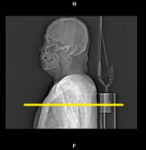
Topogram (AP)
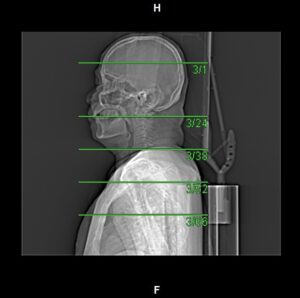
Topogram (Lateral)
Type of contrast used:
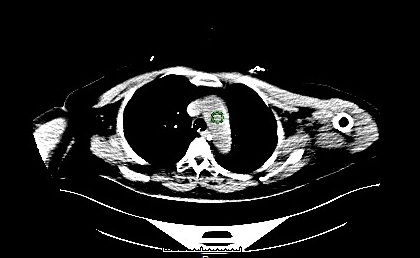
Needle Placement Test:
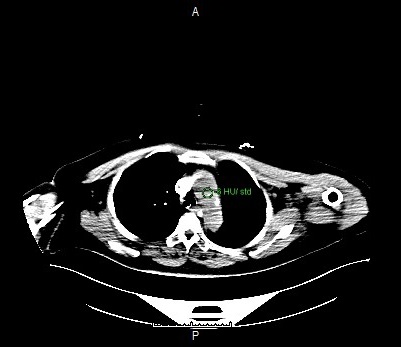
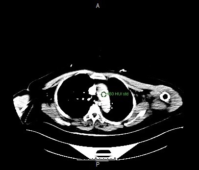
Contrast Injection:
N/A
Enter the destination URL
Or link to existing content