Skip to content
default-logo
Menu
  • Home
  • About Us
    • Background
    • Radiology Equipment
    • Webmaster & Author
  • Academics
    • Radiographic Protocols
    • Radiography Forum
    • Academic Staff Research
  • Profile
    • Lost Password
  • More
    • Microsoft Office 365
    • OT Calling Form
  • Login
  • Membership

AskRadiographer Knowledgebase

Popular Search ctamrictvcta brainct neck

CT Scan - Body Region

  • CT THORAX, ABDOMEN AND PELVIS (TAP) WITH CONTRAST
  • CT PANCREAS
  • HRCT THORAX
  • CT THORAX WITH CONTRAST
  • CT LIVER WITH CONTRAST – 3 PHASES PROTOCOL
  • CT RENAL WITH CONTRAST – 4 PHASES PROCOTOL
  • CT PELVIS NON CONTRAST
  • CT COLONOSCOPY

MRI - Spine Region

  • MRI SPINE METAL REDUCTION
  • MRI CERVICAL

MRI - Lower Limb Region

  • MRI RIGHT KNEE

MRI - Head Region

  • MRA & MRV OF BRAIN
  • MRI BRAIN – IGS PROTOCOL
  • MRI IAM
  • MRI SPECTROSCOPY

ANGIOGRAPHY - OTHERS

  • ACQUIRING IMAGES IN 3D DSA
  • ANGIOGRAPHY – IMAGE FUSION

MRI - Body Region

  • MRI SPINE METAL REDUCTION
  • MRI ENTEROGRAPHY (SMALL BOWEL STUDY)

CT Scan - Others

  • CT GUIDED INTERVENTION

Angiography - Body Region

  • SIALOGRAM

CT Scan - Head Region

  • CT FACIAL WITH CONTRAST
  • CT CERVICAL SPINE
  • CTA CAROTID ARTERY
  • CT BRAIN NON CONTRAST
  • CT BRAIN – VENOGRAPHY
  • CT BRAIN CONTRAST (IGS PROTOCOL)
  • HRCT TEMPORAL BONE NON CONTRAST
  • CTA BRAIN

CT Scan - Upper Limb Region

  • CT SHOULDER – NON CONTRAST
  • CT WRIST – NON CONTRAST
  • CT ELBOW JOINT – NON CONTRAST

Mammogram

  • Mammogram Stereotactic
  • Home
  • KB
  • Computed Tomography
  • CT Scan - Head Region
  • CT FACIAL WITH CONTRAST

Anatomy of Facial Bone

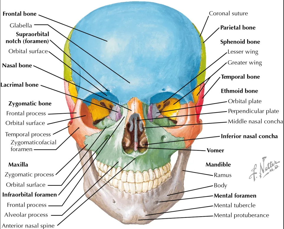
Image taken from plasticsurgerykey.com
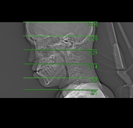
Appearance of Facial Bone in Axial CT Image

CT of Facial Bone

Examination Overview

Protocol Structure

00_FacialBone (Adult)

  • Topogram
  • Facial Bone NC

Contrast

  • Facial Bone IV

Patient Orientation Registry in System

  • Set the orientation patient in system: Head First – Supine

Topogram

  • Position the patient in head first supine position.
  • Align the patient in Mid-Sagittal plane of the table.
  • Position the transverse laser light beam at the level of 1 inch above head vertex.

Topogram Parameters

  • Topogram length: 256 cm
  • Slice: 0.6 mm
  • Scanning direction: Craniocoudal
  • Tube position: Lateral
  • Stop the topogram scanning when the scanning reach / pass over the chin

Non Contrast

  • Plan the Scan FOV (SFOV) box at topogram image
  • Set the top line at the level of head vertex.
  • Set the bottom line at the level of mental protuberance.
  • Ensure the lateral line to cover patient’s facial superficial structure (nasal bone) and skull.
  • Remind the patient to keep still during scanning.

Scanning Parameters

  • kVp:
  • mAs: Tube Current Modulation (TCM)
  • Scanning Direction: Craniocaudal
  • Scan Delay: 4 s
  • Slice:0.6
  • Image Comment: Non Contrast
  • Pitch: 0.6

Reconstruction of Non Contrast

Contrast

Type of contrast used:

  • Non-ionic iodinated contrast media
  • 300 I mg/mol

Contrast Injection:

  • Volume of Contrast Media: 50 ml
  • Volume of Normal Saline: 20 ml
  • Method of injection: Hand Bolus

Post Contrast Scan Planning

Post Contrast

  • Plan the Scan FOV (SFOV) box at topogram image
  • Set the top line at the level of head vertex.
  • Set the bottom line at the level of mental protuberance.
  • Ensure the lateral line to cover patient’s facial superficial structure (nasal bone) and skull.
  • Remind the patient to keep still during scanning.

Reconstruction of Post Contrast

Multiplanar Reconstruction (MPR)

Series of Images Send to PACS

  1. Topogram 0.6 T20f
  2. Facial Bone NC 5.0 H60s
  3. Facial Bone NC 5.0 H30s
  4. Facial Bone NC 1.0 H60s
  5. Facial IV 5.0 H30s
  6. Facial IV 1.0 H30s
  7. Patient Protocol
Do you think this article helpful?
Share This Article :
  • Facebook
  • Twitter
  • LinkedIn
  • Pinterest
Still stuck? How can we help?

How can we help?

Updated on January 20, 2023
CT CERVICAL SPINE

Quick Links

Patient Preparation

Postgraduate Research

AskRadiographer Knowledgebase

Download

About

Background

Radiology Equipment

Webmaster & Author

Sponsors

Testimonials

External Links

Radiology cases

KRIM Facebook

AskRadiographer Facebook

KOM Radiology Website

Our Location

Contact

Legal Disclaimer

Sitemap

AskRadiographer.com© Copyright 2020 - 2025. All rights reserved.
Facebook Twitter Youtube