Image taken from Microbe Notes
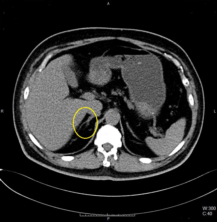
Right Adrenal Gland
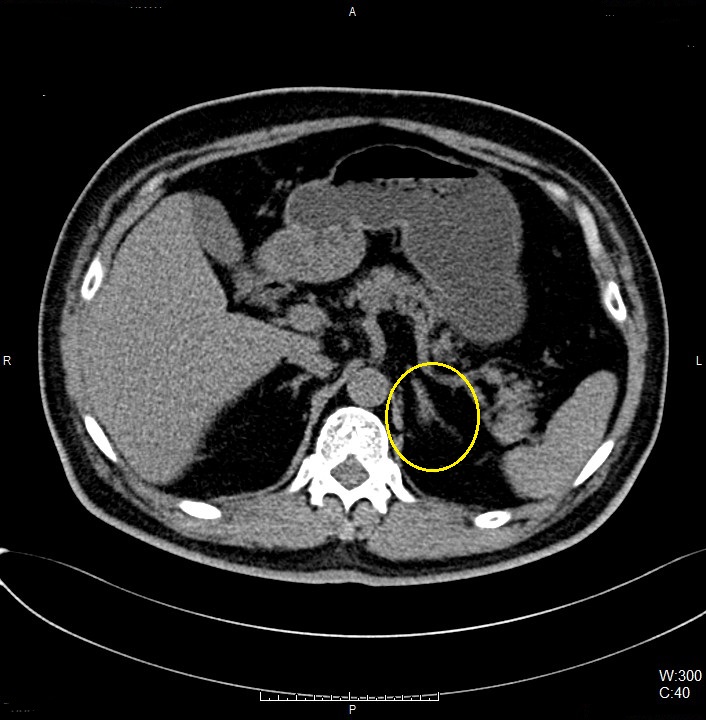
Left Adrenal Gland
Implant
Type of contrast used:
Needle Placement Test:
Contrast Injection:
N/A