Last Updated on January 20, 2023 by Mohamad Izwan

Anatomy of Thorax

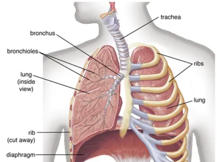
Image taken from Britannica

Appearance of CT Thorax Images

Examination Overview

Protocol Structure

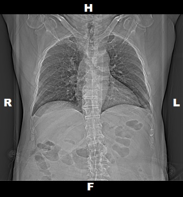
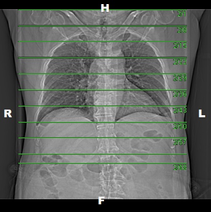
00_ThoraxRoutine (Adult)

    • Topogram
    • Non Contrast (upper shoulder to iliac crest)

Contrast

    • Portal Venous Phase
      • (upper shoulder to the iliac crest)

Topogram

  • Position the patient in head first supine position.
  • Align the patient in Mid-Sagittal plane of the table.
  • Position the transverse laser light beam at the level of patient’s upper shoulder to start the topogram.

Topogram Parameters

  • Topogram length: 512 mm
  • Slice: 0.6 mm
  • Scanning direction: Craniocoudal
  • Tube position: Top
  • Stop the topogram scanning when the scanning reach / pass over the iliac crest.

Non Contrast

  • Plan the Scan FOV (SFOV) box at topogram image
  • Set the top line at the level of upper shoulder
  • Set the bottom line at the level of iliac crest.
  • Ensure the lateral line to cover patient’s body outline.
  • Remind the patient before scanning as the breathing instruction will be given.

Scanning Parameters

  • kV: 100 kV
  • mAs: Tube Current Modulation (TCM)
  • Scanning Direction: Craniocaudal
  • Scan Delay: 4 s
  • Slice: 5.0mm
  • Image Comment: Pre-Contrast
  • Pitch: 1.2
  • Quality Reference mAs : 230

Reconstruction of Thorax Non Contrast

Contrast

Type of contrast used:

    • Non-ionic iodinated contrast media
    • 300 I mg/mol

Needle Placement Test:

    • Flow Rate: 4.5 ml/sec
    • Volume of Normal Saline: 20 ml

Contrast Injection:

    • Volume of Contrast Media:
      • 70 ml (normal body type)
      • 90 ml (large body type)
    • Volume of Normal Saline: 50 ml
    • Method of injection: Contrast Injector Single Phase

Injector setup for Normal Body Size

Injector setup for Large Body Size

Post Contrast Scan Planning

Thorax IV

  • Plan the Scan FOV (SFOV) box at topogram image.
  • Set the top line at the level upper shoulder.
  • Set the bottom line at the level of iliac crest.
  • Ensure the lateral line to cover patient’s body outline.
  • Remind the patient before scanning as the breathing instruction will be given.
  • Delay: 35 seconds.

Reconstruction of TAP IV

Series of Images Send to PACS

  1. Topogram 0.6 T20f
  2. Thorax NC 5.0 B31f
  3. Thorax NC 5.0 B80f
  4. Thorax NC 1.0 B70f
  5. Thorax IV 5.0 B31f
  6. Thorax IV 5.0 B80f
  7. Thorax IV 1.0 B31f
  8. Thorax IV 1.0 B70f
  9. Patient Protocol