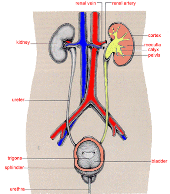
Image taken from www.le.ac.uk
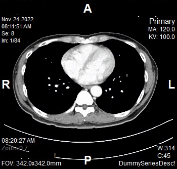
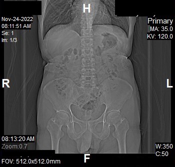
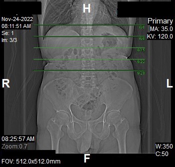
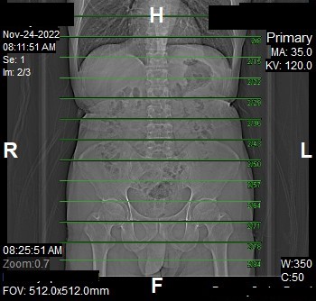
00_Renal_4Phases (Adult)
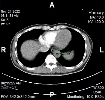
Bolus Tracking
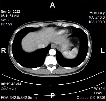
Contrast
Monitoring
Type of contrast used:
Needle Placement Test:
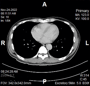
Contrast Injection: