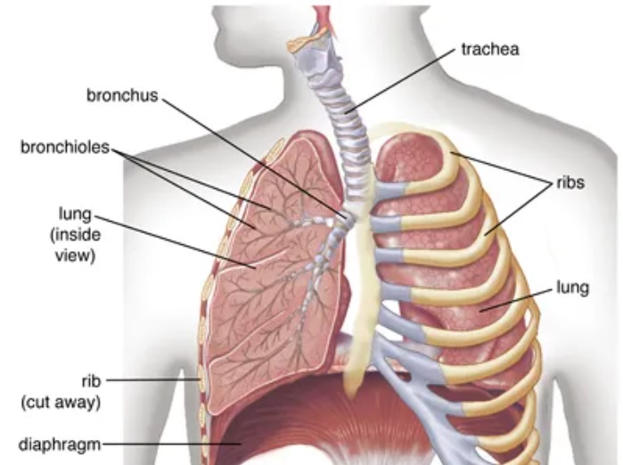
Image taken from Britannica
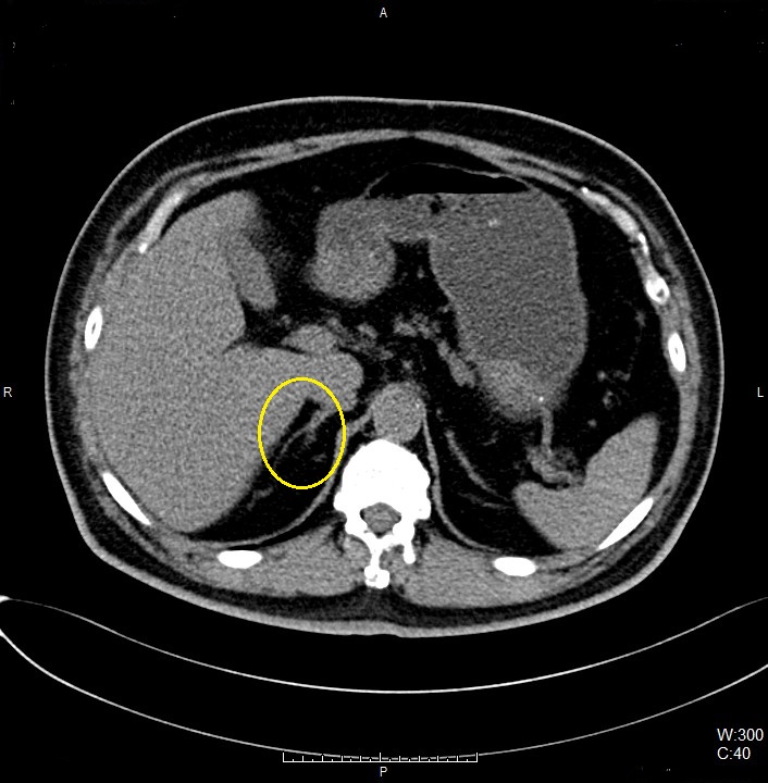
Right Adrenal Gland
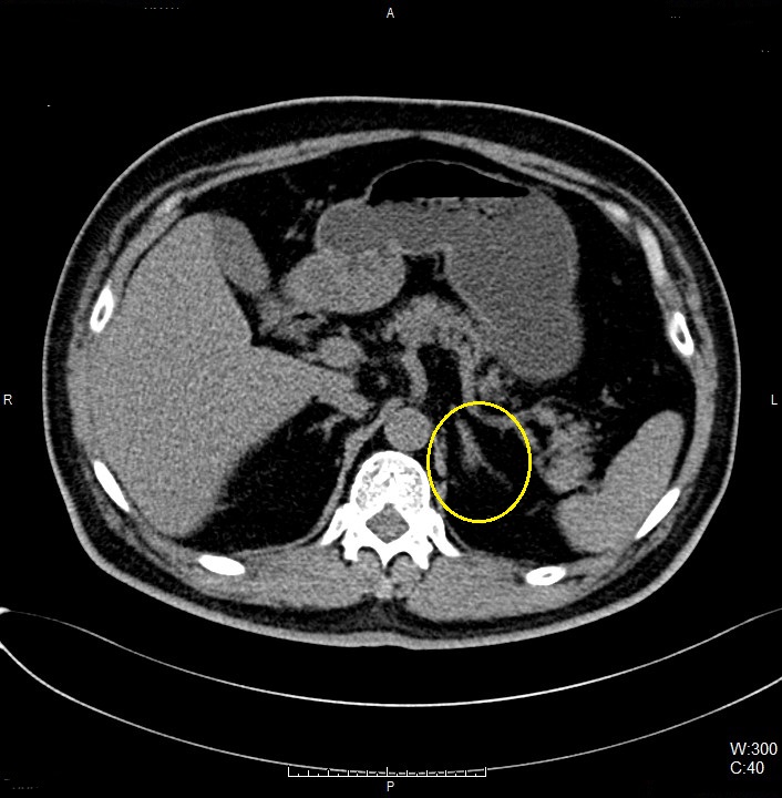
Left Adrenal Gland
00_Thorax_Veran_ENB (Adult)
N/A
Enter the destination URL
Or link to existing content