Anatomy of Wrist
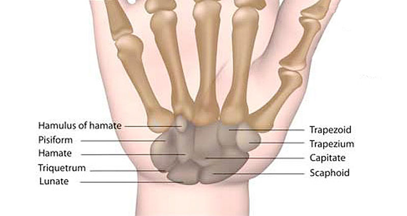
Image taken from sportsinjuryclinic.net
Appearance of Wrist in Axial CT Image
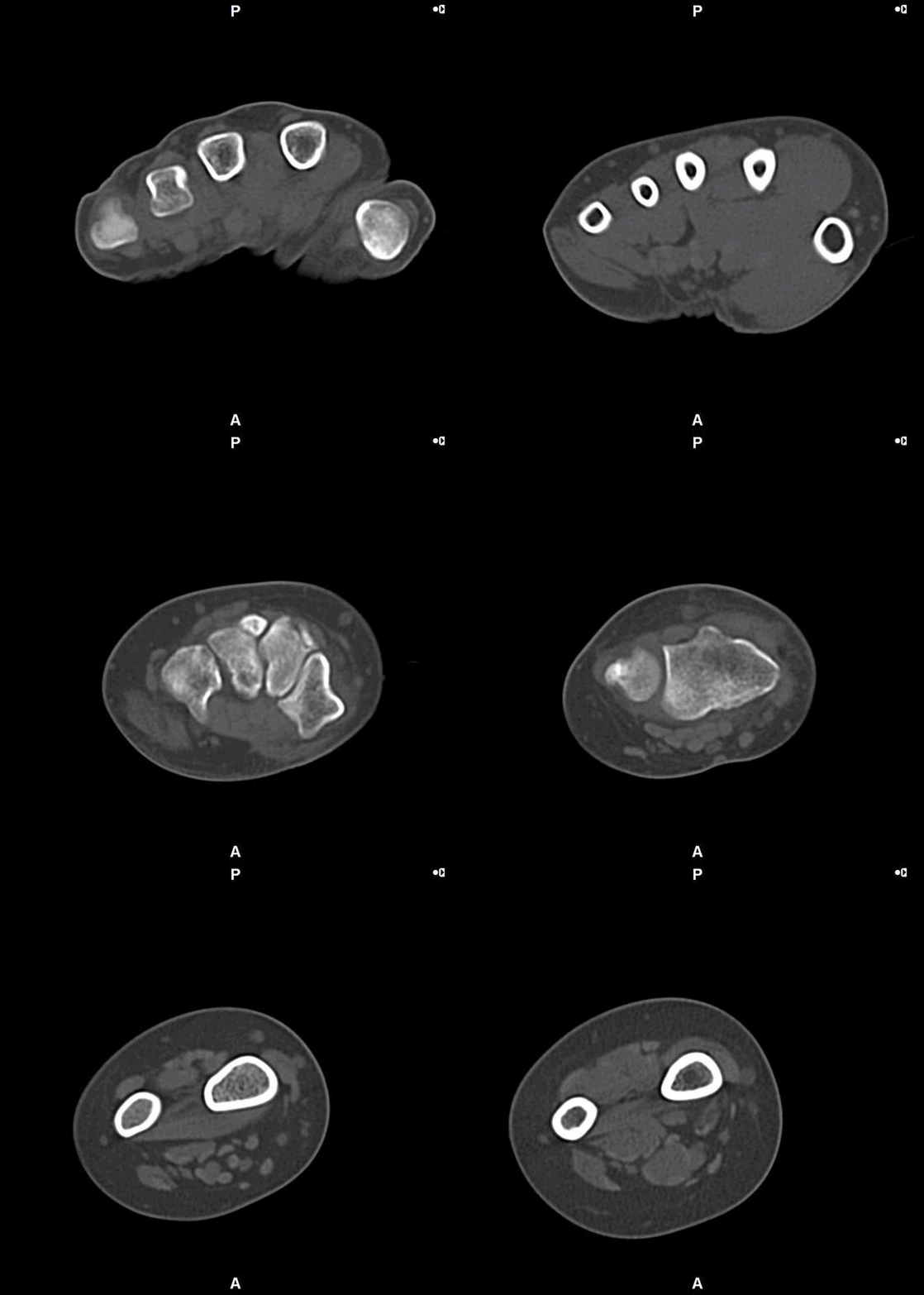
CT Wrist (Axial Images)
Examination Overview
Protocol Structure
00_Extremity (Adult)
- Topogram
- Extremity (Non Contrast)
Patient Orientation Registry in System
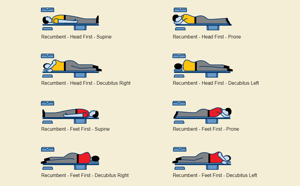
- Set the orientation patient in system : Feet First – Prone
- This is to follow the orientation of wrist (base on anatomical position)
Topogram
- Position the patient in head first prone position.
- Raise the patient’s arm above head in pronation position.
- Align the patient’s wrist in Mid-Sagittal plane of the table (if possible).
- Position the transverse laser light beam at the level of patient’s finger to start the wrist topogram.
Topogram Parameters
- Topogram length: 256 cm
- Slice: 0.6 mm
- Scanning direction: Caudocranial
- Tube position: Top
- Stop the topogram scanning when the scanning reach / pass over the elbow joint.
Non Contrast
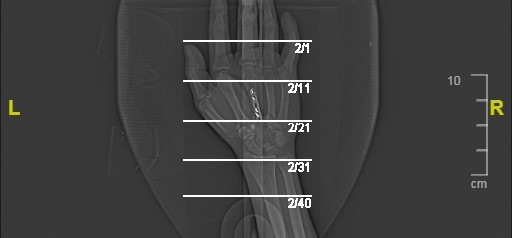
- Plan the Scan FOV (SFOV) box at topogram image
- Set the top line at the level of mid metacarpals
- Set the bottom line at the level of distal forearm (distal radius ulna).
- Ensure the lateral line to cover wrist soft tissue outline.
- Remind the patient to keep in place and not moving the arm.
Scanning Parameters
- kVp: 120
- mAs: Tube Current Modulation (TCM)
- Scanning Direction: Craniocaudal
- Scan Delay: 2 s
- Slice: 3.0mm (Acq. 128 x 0.6 mm)
- Image Comment: Non Contrast
- Pitch: 0.8
Reconstruction of Non Contrast
Series of Images Send to SyngoVia
- Extremity 1.0 B30s
Series of Images Send From SyngoVia to PACS
- Radial ranges of wrist
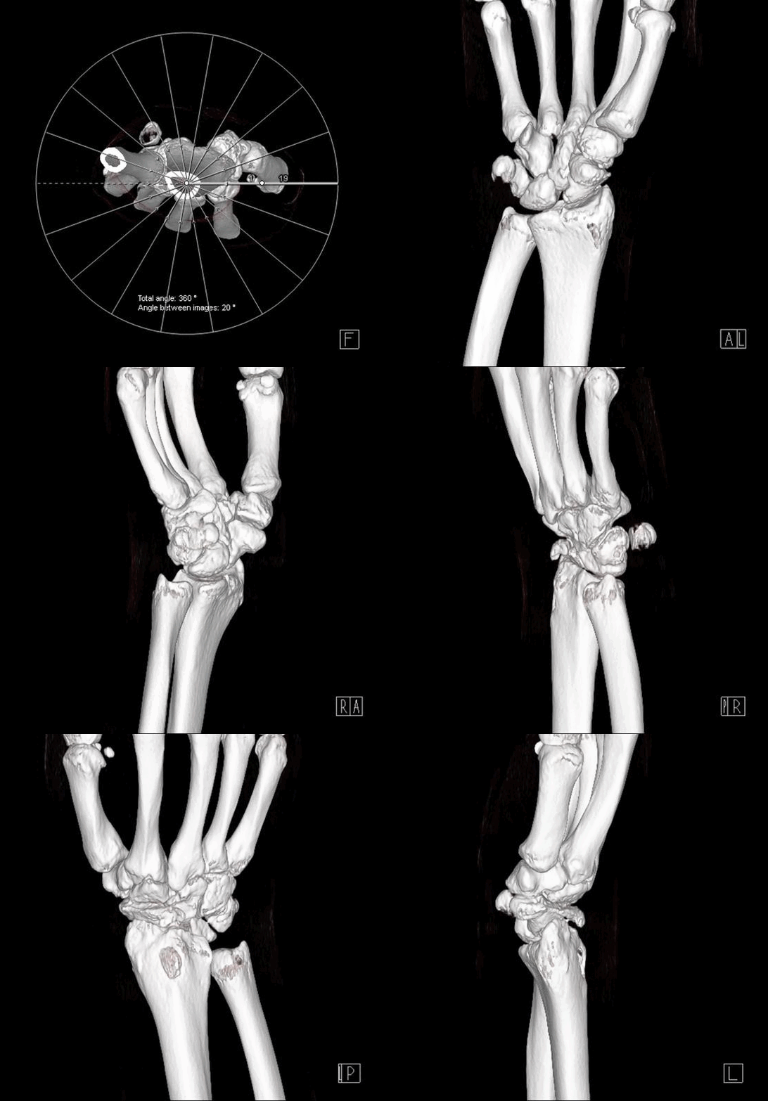
Series of Images Send to PACS
- Topogram
- Extremity 3.0 B60s
- Extremity 1.0 B60s
- Extremity 1.0 B30s


