Skip to content
default-logo
Menu
  • Home
  • About Us
    • Background
    • Radiology Equipment
    • Webmaster & Author
  • Academics
    • Radiographic Protocols
    • Radiography Forum
    • Academic Staff Research
  • Profile
    • Lost Password
  • More
    • Microsoft Office 365
    • OT Calling Form
  • Login
  • Membership

AskRadiographer Knowledgebase

Popular Search ctamrictvcta brainct neck

CT Scan - Body Region

  • CT THORAX, ABDOMEN AND PELVIS (TAP) WITH CONTRAST
  • CT PANCREAS
  • HRCT THORAX
  • CT THORAX WITH CONTRAST
  • CT LIVER WITH CONTRAST – 3 PHASES PROTOCOL
  • CT RENAL WITH CONTRAST – 4 PHASES PROCOTOL
  • CT PELVIS NON CONTRAST
  • CT COLONOSCOPY

MRI - Spine Region

  • MRI SPINE METAL REDUCTION
  • MRI CERVICAL

MRI - Lower Limb Region

  • MRI RIGHT KNEE

MRI - Head Region

  • MRA & MRV OF BRAIN
  • MRI BRAIN – IGS PROTOCOL
  • MRI IAM
  • MRI SPECTROSCOPY

ANGIOGRAPHY - OTHERS

  • ACQUIRING IMAGES IN 3D DSA
  • ANGIOGRAPHY – IMAGE FUSION

MRI - Body Region

  • MRI SPINE METAL REDUCTION
  • MRI ENTEROGRAPHY (SMALL BOWEL STUDY)

CT Scan - Others

  • CT GUIDED INTERVENTION

Angiography - Body Region

  • SIALOGRAM

CT Scan - Head Region

  • CT FACIAL WITH CONTRAST
  • CT CERVICAL SPINE
  • CTA CAROTID ARTERY
  • CT BRAIN NON CONTRAST
  • CT BRAIN – VENOGRAPHY
  • CT BRAIN CONTRAST (IGS PROTOCOL)
  • HRCT TEMPORAL BONE NON CONTRAST
  • CTA BRAIN

CT Scan - Upper Limb Region

  • CT SHOULDER – NON CONTRAST
  • CT WRIST – NON CONTRAST
  • CT ELBOW JOINT – NON CONTRAST

Mammogram

  • Mammogram Stereotactic
  • Home
  • KB
  • Computed Tomography
  • CT Scan - Upper Limb Region
  • CT ELBOW JOINT – NON CONTRAST

Anatomy of Elbow

Image taken from Microbe Notes

Appearance of Elbow in Axial CT Image

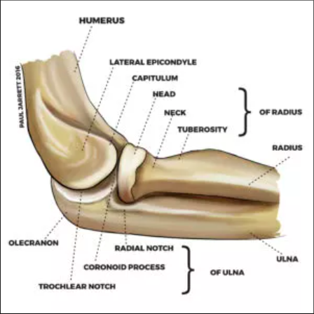
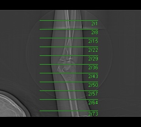
CT Elbow (Axial Images)

Examination Overview

Protocol Structure

00_Extremity (Adult)

  • Topogram
  • Extremity (Non Contrast)

Patient Orientation Registry in System

  • Set the orientation patient in system : Feet First – Supine
  • This is to follow the orientation of forearm and elbow (base on anatomical position)

Topogram

  • Position the patient in head first supine position.
  • Raise the patient’s arm above head in supination position.
  • Align the patient’s elbow in Mid-Sagittal plane of the table (if possible).
  • Position the transverse laser light beam at the level of patient’s forearm to start the elbow topogram.

Topogram Parameters

  • Topogram length: 256 cm
  • Slice: 0.6 mm
  • Scanning direction: Caudocranial
  • Tube position: Top
  • Stop the topogram scanning when the scanning reach / pass over the elbow joint.

Non Contrast

  • Plan the Scan FOV (SFOV) box at topogram image
  • Set the top line at the level of mid forearm (radius ulna)
  • Set the bottom line at the level of mid arm (humerus).
  • Ensure the lateral line to cover elbow soft tissue outline.
  • Remind the patient to keep in place and not moving the arm.

Scanning Parameters

  • kVp: 120
  • mAs: Tube Current Modulation (TCM)
  • Scanning Direction: Craniocaudal
  • Scan Delay: 2 s
  • Slice: 3.0mm (Acq. 128 x 0.6 mm)
  • Image Comment: Non Contrast
  • Pitch: 0.8

Reconstruction of Non Contrast

Series of Images Send to SyngoVia

  1. Extremity 1.0 B30s

Series of Images Send From SyngoVia to PACS

  1. Radial ranges of elbow

Series of Images Send to PACS

  1. Topogram
  2. Extremity 3.0 B60s
  3. Extremity 1.0 B60s
  4. Extremity 1.0 B30s
Do you think this article helpful?
Share This Article :
  • Facebook
  • Twitter
  • LinkedIn
  • Pinterest
Still stuck? How can we help?

How can we help?

Updated on November 17, 2022
CT WRIST – NON CONTRAST

Quick Links

Patient Preparation

Postgraduate Research

AskRadiographer Knowledgebase

Download

About

Background

Radiology Equipment

Webmaster & Author

Sponsors

Testimonials

External Links

Radiology cases

KRIM Facebook

AskRadiographer Facebook

KOM Radiology Website

Our Location

Contact

Legal Disclaimer

Sitemap

AskRadiographer.com© Copyright 2020 - 2025. All rights reserved.
Facebook Twitter Youtube